Online first
Bieżący numer
Archiwum
Najczęściej cytowane 2024
O czasopiśmie
Zespół Redakcyjny
Komitet Redakcyjny
Polityka prawno-archiwizacyjna
Kup czasopismo
Klauzula informacyjna o przetwarzaniu danych osobowych
Deklaracja dostępności
Instrukcje dla Autorów
Instrukcje dla Recenzentów
Polecamy
Kontakt
Recenzenci
2024
2023
2022
2021
2020
2019
2018
2017
2016
2015
2014
2013
Redakcja i tłumaczenia
DONIESIENIE WSTĘPNE
The influence of silver nanoparticles on the process of epithelial transition in the context of cancer metastases
1
Institute of Rural Health, Lublin, Poland (Department of Molecular Biology and Translational Research)
2
Medical University, Lublin, Poland (Department of Medical Chemistry)
3
Calisia University, Kalisz, Poland (Department of Health Sciences)
Data publikacji online: 22-12-2023
Autor do korespondencji
Magdalena Matysiak-Kucharek
Institute of Rural Health, Department of Molecular Biology and Translational Research, Jaczewskiego 2, 20-090, Lublin, Poland
Institute of Rural Health, Department of Molecular Biology and Translational Research, Jaczewskiego 2, 20-090, Lublin, Poland
Med Pr Work Health Saf. 2023;74(6):541-8
SŁOWA KLUCZOWE
breast cancermetastasissilver nanoparticlesnanotoxicologyepithelial-mesenchymal transitionMDA-MB-436
DZIEDZINY
STRESZCZENIE
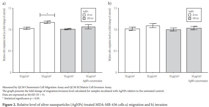
Background: Exposure to nanoparticles (NPs) can occur in a variety of occupational situations. Ultrafine particles of natural and anthropological origin toxicity has been described in epidemiological studies. Meanwhile, the risks associated with NPs exposure are not comprehensively assessed. A wide spectrum of NPs toxicity has been demonstrated, mainly through the induction of oxidative stress and inflammatory mediators. Among the newly described mechanisms of NPs toxicity is the induction of fibrosis via the epithelial-mesenchymal transition (EMT), which is also a key mechanism of cancer metastasis. The effect of NPs on EMT in the context of metastasis has not been sufficiently described so far, and the results of studies do not allow for the formulation of unambiguous conclusions. Therefore, the aim of the work was to determine the biological activity of silver NPs against MDA-MB-436 triple-negative breast cancer cells. Material and Methods: Reverse transcription real-time polymerase chain reaction was used to examine mRNA expression of EMT markers. The QCM Chemotaxis Cell Migration Assay and QCM ECMatrix Cell Invasion Assay were used to assess the level of cell migration and invasion. The tumor growth factor beta 1 (TGF-beta 1) secretion was determined by the immunoenzymatic method using the Human TGF-beta 1 ELISA Kit. Results: Silver nanoparticles (AgNPs) cause a statistically significant increase in relative expression of all tested mesenchymal EMT markers – cadherin 2, vimentin, matrix metalloproteinase 2 and matrix metalloproteinase 9. At the same time, reduction of epithelial cadherin 1 expression was observed. The level of MDA-MB-436 migration and TGF-beta 1 secretion was slighty increased in AgNPs-treated cells, with no influence on invasion potential. Conclusions: Potentially prometastatic effect of AgNPs encourages further work on the safety of nanomaterials. Med Pr Work Health Saf. 2023;74(6):541–8.
Udostępnij
Przetwarzamy dane osobowe zbierane podczas odwiedzania serwisu. Realizacja funkcji pozyskiwania informacji o użytkownikach i ich zachowaniu odbywa się poprzez dobrowolnie wprowadzone w formularzach informacje oraz zapisywanie w urządzeniach końcowych plików cookies (tzw. ciasteczka). Dane, w tym pliki cookies, wykorzystywane są w celu realizacji usług, zapewnienia wygodnego korzystania ze strony oraz w celu monitorowania ruchu zgodnie z Polityką prywatności. Dane są także zbierane i przetwarzane przez narzędzie Google Analytics (więcej).
Możesz zmienić ustawienia cookies w swojej przeglądarce. Ograniczenie stosowania plików cookies w konfiguracji przeglądarki może wpłynąć na niektóre funkcjonalności dostępne na stronie.
Możesz zmienić ustawienia cookies w swojej przeglądarce. Ograniczenie stosowania plików cookies w konfiguracji przeglądarki może wpłynąć na niektóre funkcjonalności dostępne na stronie.


